RETINAL DYSTROPHIES
Hereditary diseases related to retinal dystrophies, with the exception of pigmentary retinosis, are rare. This means that making an exact identification and diagnosis is difficult, given that there may be in the region of 70 different diseases, with very few cases of them known in the world.
- The term Retinal Dystrophies is given to hereditary retinal diseases which are more or less symmetrical and progressive.
- Retinal Degenerations are how we refer to retinal diseases which are non-hereditary, progressive, unilateral or bilateral.
Type of inheritance :
- Autosomal dominant : There are people affected in every generation. Both men and women are affected equally. The disease is transmitted solely by affected individuals. Healthy people do not transmit it. The risk of a child suffering from this disease if only one of the parents is affected is 50% with every birth.
- Autosomal recessive : only members of the same generation are affected. The parents of patients are seldom affected. It is transmitted by healthy carriers. The possibility of inheriting the affected chromosome from a parent is 50% and inheriting both affected chromosomes and therefore developing the disease is 25%. Frequently, there is consanguinity between progenitors. Men and women are affected with equal frequency.
- Recessive linked to gender : Only men suffer from the disease. Women are all carriers. The odds of having an affected child by a mother who is a carrier are 50% and their daughters also have a 50% chance of being carriers themselves. The affected gene is found in the X chromosome.
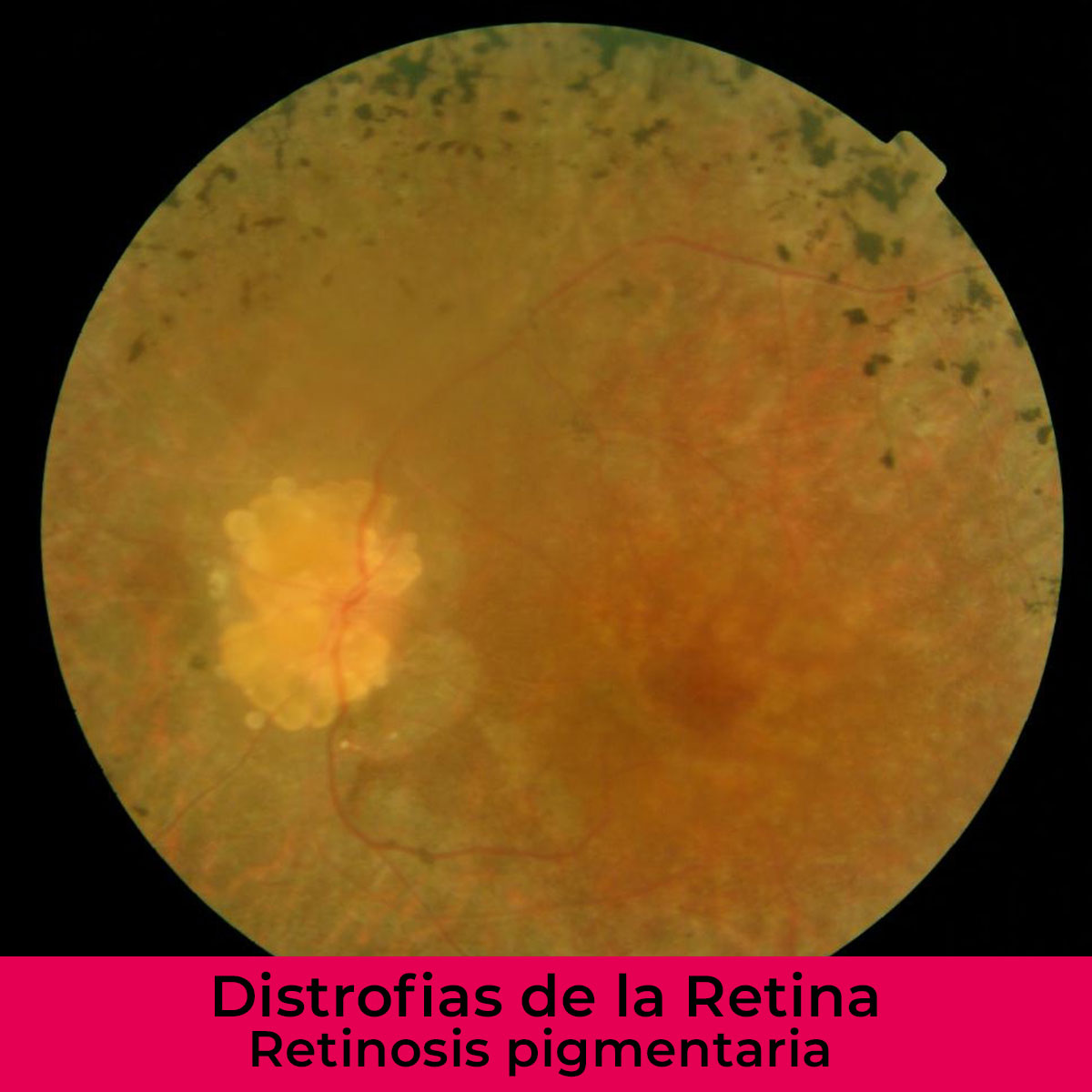
PIGMENTARY RETINOSIS
This is a group of hereditary retinal diseases which are characterized by : clinically bad night vision , progressive constriction of the visual field and a progressive loss of visual acuity. On examination of the fundus of the eye characteristic lesions called bone spicules are to be found and there is also a reduction in blood vessels, paleness of the eyelids and macular edema.The inheritance may be autosomal dominant , recessive or linked to gender. There are sporadic cases which are non- hereditary. There is a better prognosis with dominant types. They usually begin between the first and third decade of life and have a slow progress, with periods where the condition worsens.
Pigmentary Retinosis can present in a variety of ways, both in symptoms and in the appearance of the fundus of the eye. There are different clinical types, such as PR without pigmentation, unilateral PR, inverse PR, pericentral PR…It can also be found associated with other congenital pathologies within general syndromic patterns. The pathology finding is the progressive degeneration of photoreceptors which are initially rods , latterly there are also cones and the internal layers of the retina are together with the pigmentary epithelium. In the final stages there is a proliferation of glial cells in the retina and the optic nerve with hyalinization of the vessels.
STARGARDT DISEASE
This is also known as juvenile macular degeneration. It is a hereditary disease in which the loss of cones predominates and therefore a loss of day vision ensues. It forms part of a group of retinal dystrophies which are called Bull's eye maculopathy. This is clinically characterized by a progressive loss of visual acuity and bad color vision. Occasionally there is also photophobia. When examined, the macula displays the characteristic appearance of beaten metal when the disease has evolved, although it can appear normal at the outset.
Autosomal recessive is inherited.
It usually begins between the first and second decade of life.
The pathology study reveals a complete loss of pigmentary epithelium and photoreceptors in the macula.
CONE / ROD DYSTROPHIES
There are a series of hereditary retinal diseases that share symptoms with pigmentary retinosis and Stargardt disease. In these it is fundamentally the cones which are affected but progressively the rods can be too. Globally they are known as cone/ rod dystrophies. In these we can find a decrease in visual acuity along with an alteration in the perception of colors. This is accompanied by a constriction of the visual field and bad adaptation to darkness.
There are also retinal dystrophies which predominantly affect the visual field which differ in the way they present from the classic case of Stargardt disease and which we call cone dystrophy.