SPECULAR MICROSCOPY
This is the study of the endothelial cells in the cornea.
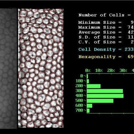
Specular microscopy study of the corneal endothelium
The corneal endothelium is the layer of cells which is found in the (deep) posterior face of the cornea and which is fundamental for the cornea to function. A decrease in the number of endothelial cells may cause a loss of transparency in the cornea, with a subsequent decrease in vision.
This test is asked for whenever the possibility of using an intraocular lens for refractive purposes is recommended or when cataract surgery is advised.
With specular microscopy an endothelial count based on surface area can be calculated, thereby determining whether there is any change in the shape or size of the endothelial cells.
It should be noted that there is a natural loss of cells related to age. Endothelial cells do not reproduce, with the result that when there is a significant decrease in their number , the possibility of a corneal transplantation to give the cornea back its natural transparency and therefore restore vision is to be considered.