KERATOCONUS
Keratoconus is suspected when a young person with elevated astigmatism and myopia does not achieve good vision with glasses and where there are frequent changes in graduation.
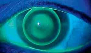
Corneal topography confirms the diagnosis.
What is Keratoconus ?
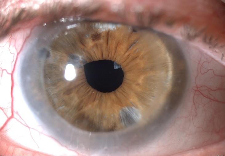
Keratoconus is a progressive deformity of the cornea, which adopts an irregular conical shape due to the alteration of the internal structure of the corneal tissue. It normally appears between the ages of 10 and 20, with a deterioration in vision caused by myopia and progressive astigmatism. Each eye can be affected although the evolutionary degree may be different. The cornea becomes thinner and more deformed as the keratoconus develops, which increases the level of astigmatism. This can rarely be treated by using glasses. Corneal topography confirms the diagnosis and also gives us the information needed to carry out treatment.
What are the causes ?
In keratoconus there exists a weakening of the cornea due to an alteration in the collagen fibers which it is made up of, but the actual cause is uncertain. There is a hereditary pattern in some patients, but the majority of patients do not have a family history of it. Another possible cause is the constant rubbing of eyes, as there exist a greater number of patients with keratoconus who suffer from allergies. Keratoconus occurs more frequently in patients who have diseases where there is an alteration of collagen in different parts of the organism (Down syndrome, Marfan, mitral valve prolapse etc…)
How is it treated ?
For many years, professionals who are specialists in keratoconus have had very limited options for treating this condition . The use of rigid contact lenses or more recently the use of Intrastromal rings to help alleviate problems with vision have been the only tools at their disposal, before arriving at the situation of performing a corneal transplant. At the Hoyos Ophthalmological Institute we have incorporated Corneal Crosslinking into our treatment of keratoconus, which is the most advanced technology and in which we carry out a procedure which reinforces the weakened cornea by creating new bonds of molecular collagen , thus attempting to detain the advance of the disease and with it the accompanying deterioration in vision.
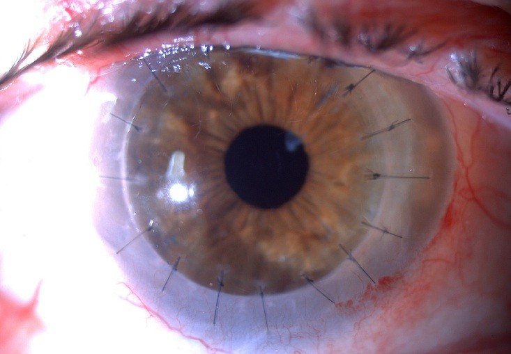
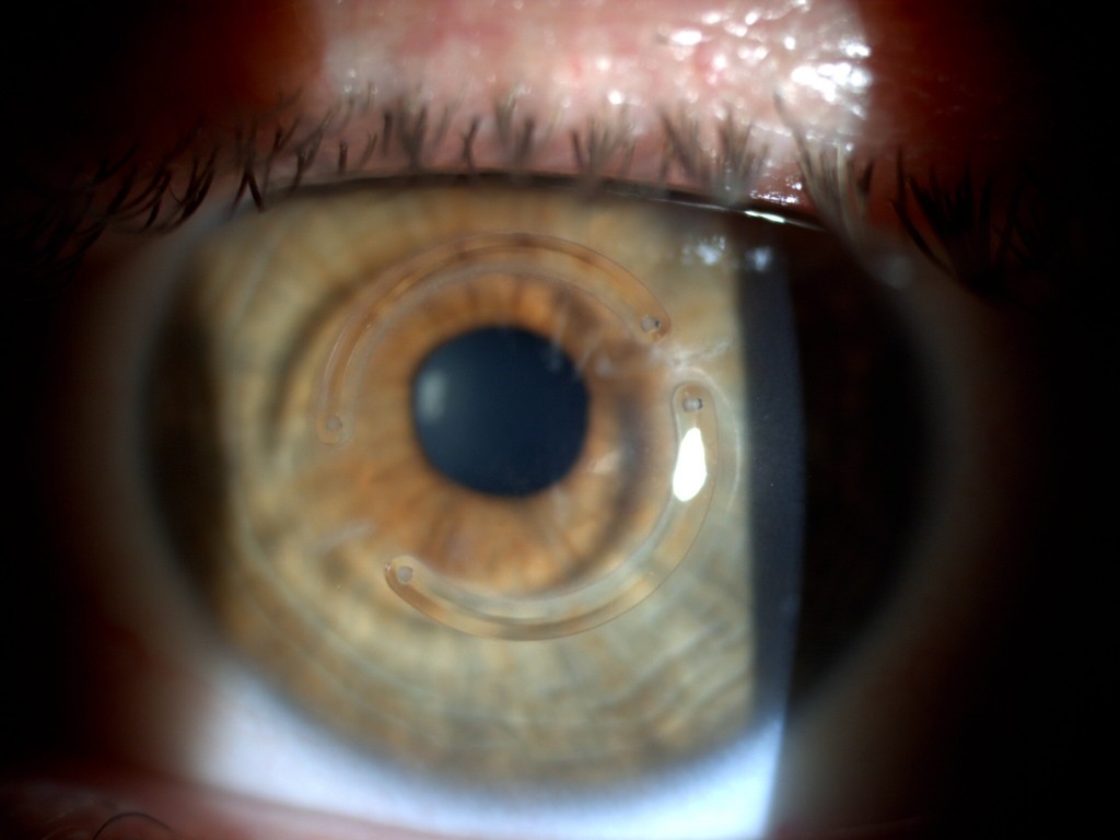
Implanting Intracorneal rings is a surgical treatment in which two semi-circular segments are implanted in the interior of the cornea, in an attempt to correct the corneal irregularity which conforms to keratoconus , thereby improving as much as possible the patient’s eyesight. With a corneal transplant, the objective is to substitute the central part of the patient’s cornea, replacing it with a donor cornea. Transplantation is only advisable for advanced cases in which the patient cannot achieve good vision by wearing contact lenses and where the placing of rings or other treatments is not indicated.
Corneal Crosslinking with riboflavin is a new method for treating progressive keratoconus as well as any other variant of corneal ectasia. The treatment consists in scraping the surface of the cornea to apply riboflavin, a substance which sensitizes the collagen. Then the creation of these new bridges or unions between the collagen fibers is stimulated, with the application of an ultraviolet light for a period of 30 minutes.