OCT- OPTICAL COHERENCE TOMOGRAPHY
OCT- Optical Coherence Tomography is nowadays an indispensable tool in the practice of ophthalmology. It is a technique which is used for diagnosis, checking and follow-up, which allows us to study live histological sections of both the retina and cornea.This technology is based on a complex optical principle named interferometry.
OCT- Optical Coherence Tomography is a technique which is painless for the patient, fast, extremely reliable and reproducible.
OCT- Optical Coherence Tomography is used :
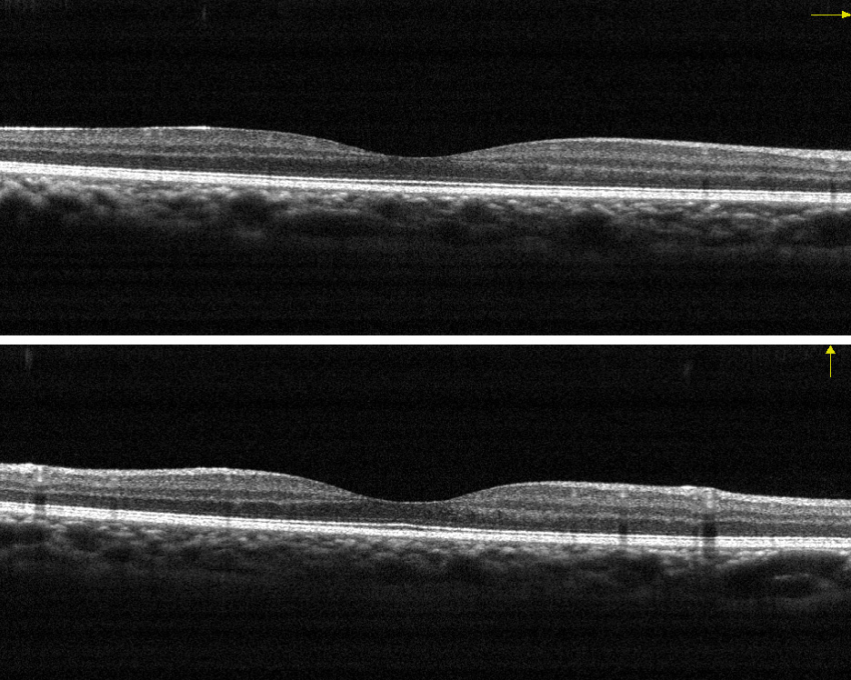
- In the study of the macula, the structure of the retina which is responsible for maximum visual acuity. OCT allows us to obtain histological sections of the retina , from the anterior to the posterior face and analyze them microscopically, thereby permitting us to observe the pathological changes which are produced. This is a fundamental test when following up cases where patients who have macular degeneration associated with age (AMD), diabetic macular edema, macular edema secondary to thrombosis, macular holes, epiretinal membrane, vitreomacular traction, central serous choroidopathy etc..
- With glaucoma, OCT- Optical Coherence Tomography allows us to study the head of the optic nerve and the layer of nerve fibers , thanks to its great precision, reliability and reproducibility. It enables us to make a diagnosis, and what is most important in this pathology, it allows us to keep a check on the damage which is produced over time with glaucoma.
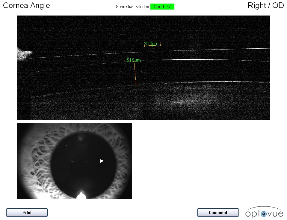
- As for the cornea, OCT- Optical Coherence Tomography permits us to carry out complete sections of the cornea, enabling us to obtain corneal pachymetry and to study scars and their depth.