RETINOGRAPHY
Photography of the eye’s fundus.


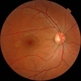
Retinography is the photographic study of the retina. It is a technique which is used for the diagnosis and follow-up of pathologies relating to the fundus of the eye.
The retinographer takes a detailed digital photograph of the fundus of the eye, with a resolution of between 2 and 5 megapixels, therefore making it highly unlikely that any anomaly is overlooked in this test.
In addition to conventional non- mydriatic retinography, the H.O.I. has Compass retinography which allows us to achieve high quality images of the fundus of the eye of up to 60º and which, thanks to Confocal True Color image technology, permits us to obtain the true color of the retina, even through pupils of 3mm in diameter
Photographs of the eye’s fundus provide us with graphic documentation of the changes the retina undergoes of patients with diabetes and arterial hypertension and are of use to us to find out and adapt metabolic checks on these diseases in each case. Moreover, we are able to photograph internal tumoral lesions to control their growth and also any vascular or degenerative pathology of the retina. Photographic checking of the head of the optic nerve in a patient with glaucoma allows us to be aware of any evolution in the process of atrophy of the optic nerve.