RETINA AND VITREOUS
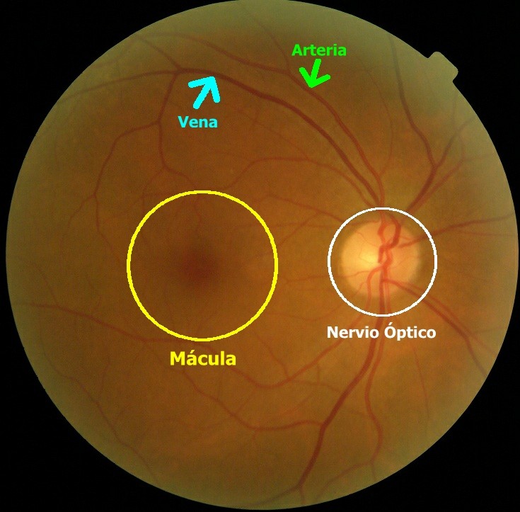
The retina is the layer of light sensitive tissue which is found in the internal posterior part of the eye and which acts like the film in a photographic camera : when signals of light enter the eye, they pass through the cornea and the crystalline lens and are focused on the retina. The retina is formed by nerve cells which convert light into electric signals and then sends them through the optic nerve to the brain, where they are converted into visual images. The optic nerve goes from the brain to the center of the retina approximately and then it branches out. The retina is normally red in color due to the rich blood flow which it receives. The central part of the retina, which is called the macula, contains the greatest density of light sensitive nerves and consequently produces the best visual resolution. The retinal vein and artery reach the retina near the nerve, where they branch out, following the route of the nerves. As well as the optic nerve and its ramifications, the retina also has a great number of vessels which carry blood and oxygen. (SEE PHOTOS)
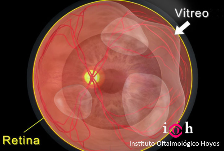
The vitreous looks very similar to a transparent egg white and fills the central cavity of the eye. The vitreous is attached to the retina, principally in the posterior part of the eye, the optic nerve, the macula and the large retinal blood vessels. (SEE PHOTOS)
Anyone who experiences changes in their perception of clarity or color, flashes of light, floaters or distorted vision should have their retina examined.